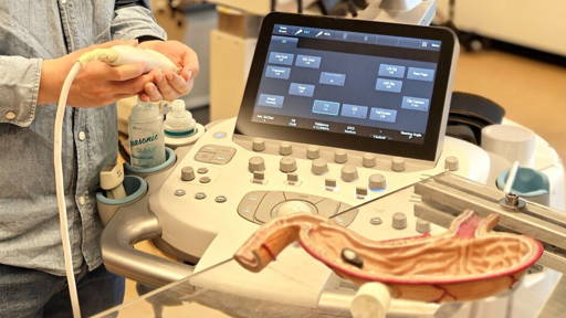
German scientists have succeeded in using 3D technology to map and identify tumor environments to determine whether a patient may benefit from personalized cancer treatment. When determining a treatment, not only an understanding of the type of cells in a tumor is important. How these cells interact with cells in their environment also plays an important role. With their research, potential targets for personalized cancer therapy can be identified, according to the study.
The international research team led by the Berlin Institute for Medical Systems Biology at the Max Delbrück Center (MDC-BIMSB) combined 3D spatial transcriptomics and extracellular matrix imaging to obtain extraordinary details about the inner workings of an early-stage lung tumor. The proof-of-concept study was published in Cell Systems.
“Tumors are complex ecosystems where tumor cells are in close contact with the surrounding extracellular matrix. They interact with many other cell types. With the new 3D mapping technology, the data we can now obtain in a patient's tumor tissues becomes so accurate and comprehensive that we can computationally predict the molecular mechanisms that drive phenotypes. This is new and of fundamental importance to make personalized medicine a reality,” said Professor Nikolaus Rajewsky, MDC-BIMSB director, head of the Systems Biology of Regulatory Elements laboratory.
From 2D to 3D mapping
Transcriptomics documents which RNA expression is actively expressed in cells, indicating which activities the cell is performing and which cell types are present in a sample. Spatial transcriptomics does this, but for individual cells, to create a 2D map.
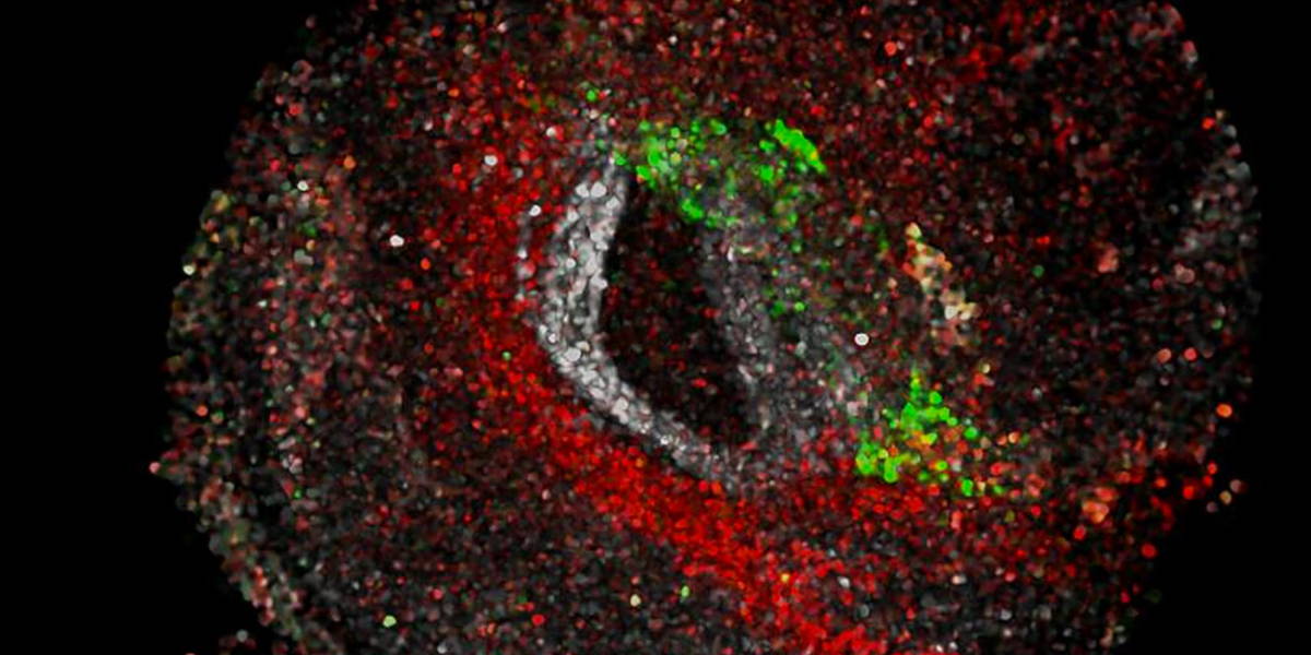
Early in its development, the research team gained access to a CosMx instrument from the company NanoString, which allows this mapping to be performed at extremely high resolution. Thus, up to 1,000 different RNA molecules can be detected simultaneously, compared to traditional methods that identify only a handful of molecule types at a time. In this way, the team analyzed 340,000 individual cells from the lung tumor and identified 18 cell types.
The 3D analysis was driven by a new computational algorithm, STIM, which aligns datasets to reconstruct virtual 3D tissue blocks. “We realized that datasets from spatial transcriptomics can be modeled as images,” said Dr. Nikos Karaiskos, postdoctoral researcher in the Rajewsky lab. The operation of STIM is described in detail in Cell Systems.
Imaging technique
The researchers then collaborated with the Systems Biology Imaging Platform in Berlin to apply a separate imaging technique, called second harmonic generation, to map elastin and collagen to cellular environments, which in the lung are the main components of the extracellular matrix. Areas with more elastin were healthier, while areas with more collagen surrounded tumor cells, indicating harmful tissue remodeling.
“So not only do we know which cell types are present, we also know how they are grouped relative to their neighbors, and we could begin to understand how tumor cells restructure non-malignant cells at the tumor surface to support tumor growth,” explained Tancredi Massimo Pentimalli, MD who is a PhD researcher.
Communication between cells
Ultimately, the team was able to understand precise phenotypes - for example, whether fibroblasts, which form connective tissue, were activated and remodeling the tissue or not. They were also able to listen to communication between cells and determine how tumor cells blocked immune cells from entering the tumor.
“This immunosuppression mechanism is known and suggests that immunotherapy could help,” he said. Immune checkpoint inhibitors would reverse the suppression and then you have an army of immune cells already ready to attack. It was exciting to see how our approach identified these dynamics and could help determine a personalized immunotherapy plan,” said Pentimalli
Remarkably, these important insights were only possible with 3D data - in 2D, it was impossible to distinguish between the tumor and other immune cells embedded in the tumor surface.
Routine tissue samples
Although this new 3D mapping technology is particularly high-tech, it all starts with a routine tissue sample found in any pathology lab. For their research, the scientists used a tissue sample from a lung tumor that was several years old, preserved with formalin and embedded in kerosene wax - the standard method pathologists use to preserve archival tissue.
“We were able to extract all this wealth of molecular information from a very thin piece of once sample that had been sitting at room temperature for years.This is pathology 2.0: not just looking at cells under a microscope to make a diagnosis, but bringing molecular insight to the clinic,” Pentimalli said.
Now that the proof-of-concept has been established, the team plans to apply the approach to larger data sets. They are currently working on 700 samples from 200 patients and are collaborating with Dr. Fabian Coscia, head of the Spatial Proteomics Lab at the Max Delbrück Center, to incorporate protein activity into the analysis.