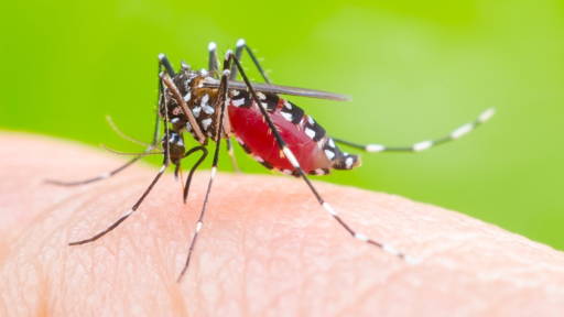
An AI-supported analysis tool helps pathologists to assess tissue samples from skin cancer patients more consistently. This is the conclusion of an international study led by the Karolinska Institutet, in collaboration with Yale University. By using AI to interpret tumour-infiltrating lymphocytes (TILs), important biomarkers in melanoma and other cancers, pathologists were able to more reliably assess how aggressively the tumour is developing.
The use of AI not only increased the reproducibility of the assessments, but also improved prognostic accuracy. TILs are immune cells that occur in or around tumours and provide information about the body's immune response. In malignant melanoma, the presence of these cells plays a crucial role in both diagnosis and treatment strategy. Because estimating the number of TILs is currently subject to subjective interpretation, it is a vulnerable link in pathological practice. The AI tool tested in this study offers a solution by objectively quantifying the number of TILs based on digital microscopy images.
Relatively small study
The study involved 98 pathologists and researchers from other professions, who were divided into two groups. One group consisted exclusively of experienced pathologists. They worked according to standard procedures. That is to say, they viewed digital images of stained tissue sections and estimated the number of TILs according to current guidelines. The second group consisted of pathologists, but also researchers from other professions, all of whom had some experience in assessing pathological images.
They also viewed the images according to standard practice, but were assisted by AI support that quantified the number of TILs. Everyone assessed 60 tissue sections, all from patients with malignant melanoma. The study was retrospective, so the images showed tissue samples from patients whose diagnosis and treatment had already been determined.
The results, published in JAMA Network Open, speak for themselves: pathologists who used AI were much more consistent in their assessments. In addition, their prognoses were found to be more in line with the actual clinical outcome of the patients, confirming that AI can be a valuable tool in risk stratification and treatment decisions.
Although further validation is needed before this technology can be widely applied in clinical practice, researcher and pathologist Balazs Acs emphasises that the results are promising. ‘AI can objectify the assessment process and thus contribute to safer, personalised care for cancer patients.’
AI and medical diagnostics
It is no longer news that AI applications can take medical diagnostics and pathology to the next level. Last year, a pilot was conducted in the United Kingdom with an AI platform capable of assessing within seconds whether a skin lesion is malignant or not. And earlier this year, an AI model capable of analysing long-term ECGs was tested for an international study. With the help of the AI tool, the number of missed diagnoses decreased by a factor of 14.
In the Netherlands, too, people are working hard on AI solutions that can contribute to earlier and more accurate diagnoses. For example, in 2022, scientists at Maastricht University developed an AI method that can better estimate the size of a tumour so that the possible follow-up course of radiation treatment can be determined more accurately.