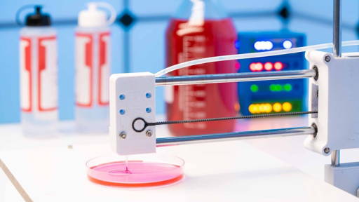
Researchers from UCL and the Francis Crick Institute have identified, in a living mouse embryo, the origin of heart cells for the first time using 3D images of the formation of a heart. For the study, the team used a technique called advanced light sheet microscopy on a specially developed mouse model. This is a method that uses a thin sheet of light to illuminate tiny samples and take detailed pictures, creating clear 3D images without damaging living tissue.
This allowed them to follow individual cells as they moved and divided over the course of two days - from a critical stage of development called gastrulation to the point where the primitive heart begins to take shape. This allowed the researchers to identify the cellular origin of the heart. Gastrulation is the process by which cells begin to specialize and organize into the body's primary structures, including the heart. In humans, this occurs during the second week of pregnancy.
Understanding congenital heart defects
The findings of the study, published in The EMBO Journal, could revolutionize the way scientists understand and treat congenital heart defects, the researchers said. “This is the first time we have been able to watch heart cells so closely and for so long during mammalian development. We first had to reliably grow the embryos in a dish for long periods of time, from a few hours to a few days, and what we found was totally unexpected,” says Dr. Kenzo Ivanovitch of the UCL Great Ormond Street Institute of Child Health and British Heart Foundation Intermediate Research Fellow.
The insights from the study could revolutionize the way scientists understand and treat congenital heart defects, which affect nearly one in 100 babies. The findings could also accelerate advances in growing heart tissue in the lab for use in regenerative medicine. “In the future, we hope this work will reveal new mechanisms of organ formation. This will lead to design principles for accurately programming tissue patterns and shapes for tissue manipulation,” Ivanovitch said.
Fluorescent markers
Using fluorescent markers, the team marked heart muscle cells (called cardiomyocytes), causing them to light up in different colors. Combined with light microscopy, this innovation allowed the researchers to create a detailed time-lapse video. Snapshots were taken every two minutes over a 40-hour period, creating images with unprecedented spatial resolution.
The resulting images showed how cells move, divide and form the first parts of an embryo, such as the heart. Each illuminating cardiomyocyte could then be traced to earlier cells, allowing scientists to create a genealogy of the cells. This allowed them to see exactly when and where the first cells making just the heart appeared in the embryo. Watch the UCL video below:
In the very earliest stages, embryonic cells were multipotent (capable of becoming different cell types). These included not only heart cells but also others such as endocardial cells, a cell type lining the inside of blood vessels and heart chambers.
Organized movement of cells
However, the researchers found that early in gastrulation (usually within the first four to five hours, after the first cell division) cells that contribute only to the heart quickly emerge and behave in a highly organized manner. Instead of moving randomly, they follow different paths - almost as if they already know where they are going and what role they will play, whether they contribute to the ventricles (the heart's pumping chambers) or the atria (where blood enters the heart from the body and lungs).
“Our findings show that the determination of the fate of the heart and the direction of cell movements are regulated much earlier in the embryo than current models suggest. This fundamentally changes our understanding of heart development by showing that what appears to be chaotic cell migration is governed by hidden patterns that ensure the proper formation of the heart,” Ivanovitch said.