Researchers at the University of British Columbia Okanagan (UBCO) have taken an important step towards personalised lung research and therapy. They have developed an advanced 3D-printed lung model that closely resembles real lung tissue. This innovation could become a game changer in the diagnosis and treatment of complex respiratory diseases such as asthma, COPD and lung cancer.
The bio-printed lung tissue was developed by a team led by Dr Emmanuel Osei, assistant professor at the Irving K. Barber Faculty of Science and affiliated with the Centre for Heart Lung Innovation. According to Osei, the new model accurately mimics the structure of the human lung. This creates a reliable research environment in which cellular responses to cigarette smoke or medication, for example, can be studied in detail. ‘Developing a realistic lung model is essential for better understanding disease mechanisms and testing new treatment methods. This model also allows us to identify new drug targets more quickly and efficiently,’ says Dr Osei.
Technological innovation
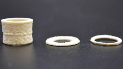
The researchers used a specially developed bio-ink consisting of light-sensitive, polymer-modified gelatin and polyethylene glycol diacrylate. This was used to print a hydrogel in which different cell types and channel structures are integrated, mimicking the blood vessels and airways of the lung. Once printed, this model exhibits similarities to the complex mechanical behaviour of lung tissue.
What distinguishes this model from previous attempts is the addition of vascular components. This also allows blood flow to be mimicked, which is essential for studying inflammatory responses, cancer progression or fibrosis formation. This step makes it possible to create a more realistic simulation of the human lung environment, with important implications for drug research.
From patient to lab
An important advantage of this technology is the ability to isolate lung cells from patient tissue and multiply them via bioprinting. Instead of waiting for new donor material, researchers can immediately start creating test models. This increases efficiency and contributes to the realisation of personalised treatments.
‘In many cases, only limited lung tissue is available for research. Thanks to this technique, we can create multiple research models from a single small piece of lung tissue,’ explains Osei.
Clinical relevance demonstrated
The clinical applicability was demonstrated by exposing the 3D model to cigarette smoke extract. An increase in pro-inflammatory cytokines was observed. These are signals that indicate an inflammatory response. These results emphasise that the model is capable of accurately simulating disease processes.
Future applications could focus on personalising treatments, testing new drugs and shortening development cycles. ‘The fact that our model can be adapted with patient-specific cells makes it ideally suited for personalised medicine,’ says Osei.
The study, published in Biotechnology and Bioengineering, was conducted with support from Mitacs and Providence Health Care. The research team is already in talks with biotech partners and other scientific consortia to explore other clinical applications.
3D biotechnology
Last year, TU/e investigated the use of advanced light printing technology (xology) with 3D bioprinting to create both healthy and diseased tissue in a single model. This new platform, called STOMP, produces tissues that can be easily removed from the device without damage. STOMP uses a mini device and capillary action to form cells and hydrogel into a free-hanging structure. This method makes it possible to combine different cell types within a single model.
By linking the platform to existing biofabrication methods, a powerful, multidisciplinary platform for biomedical research is created. It makes personalised and reproducible research into disease models accessible to more laboratories and accelerates the development of new therapies.