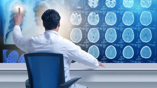
A team of Chinese scientists has achieved a major breakthrough in the field of 3D imaging of large-scale biological tissues. They claim to have developed the world's fastest high-definition 3D imaging technology for the entire body of small animals with subcellular resolution, enabling the fine architecture of the peripheral nervous system (PNS) to be mapped efficiently.
For a long time, knowledge about the architecture of the PNS was based on anatomical studies with a resolution of millimetres. Over the past decade, advances in 3D optical microscopy have led to mesoscopic connectomic mapping of the entire brain with a resolution of microns, but similar analyses for the PNS throughout the body remain a challenge.
IoT of the body
The PNS acts as the “Internet of Things (IoT)” of the body and mediates bidirectional communication and modulation between the brain and the organs. The PNS controls and regulates crucial functions such as breathing and heart rate. It also processes sensory signals such as pain and temperature. This enables efficient physiological coordination between different tissues and organs.
Mapping the intricate connections of the PNS throughout the body is essential for a fundamental understanding of the complex functional mechanisms and associated disease pathogenesis.
Existing imaging techniques struggle to find a balance between image resolution and speed. Even when combined with techniques for clearing whole-body samples, it remains a challenge to map the long, complex and often intertwined nerve pathways of the PNS on a whole-body scale.
3D imaging developments
The team previously developed volumetric imaging with synchronised on-the-fly scanning and readout technology (VISoR) for 3D imaging of parts of the brain. VISoR has the advantage of high imaging speed, high resolution and scalability. With this technology, it was already possible to image the entire brain of a mouse within 1.5 hours with a resolution of less than one micron.
However, this approach was not suitable for whole mouse bodies. Unlike the relatively compact and homogeneous brain, the body of mammals is much larger and highly heterogeneous, with irregular structures and diverse tissue types. This often leads to deformation and damage to body parts prior to VISoR imaging, making complete reconstruction difficult.
Innovative new strategy
To address these challenges, the team developed an innovative new strategy: ‘in situ cutting and 3D blockface imaging’. They developed a so-called blockface VISoR imaging system that integrated various technologies.
Based on this strategy, the researchers set up an optimised technical pipeline and achieved uniform 3D imaging with subcellular resolution of a fully adult mouse body within 40 hours, generating approximately 70 terabytes of data per fluorescence channel. In total, more than 4 petabytes of raw data from dozens of mice were collected.
The researchers explained that this new technology not only helps establish a new paradigm for mapping PNS connectivity and resolving fundamental questions about neural regulation, but also provides valuable insights into broader fields such as developmental biology, comparative anatomy, and biomedical research in general.
Room for further improvement
Furthermore, there is still room for improvement and optimisation of this technology. The next steps include using two or more cameras for efficient multi-channel imaging and investigating its application in imaging other, larger biological samples.
The team was led by Professors Guo-Qiang Bi and Pak-Ming Lau from the Hefei National Research Centre for Physical Sciences at the Microscale and the Department of Life Sciences and Medicine at the University of Science and Technology of China (USTC), in collaboration with the Institute of Artificial Intelligence, Hefei Comprehensive National Science Centre (IAI) and the Shenzhen Institute of Advanced Technology, Chinese Academy of Sciences (SIAT). The team's findings have been published in Cell.