An innovative discovery by Australian researchers could improve the treatment of hereditary heart diseases in both children and adults. Scientists at the Cardiac Bioengineering Lab at the QIMR Berghofer Medical Research Institute have used stem cell technology to develop so-called cardiac organoids. These are “mini hearts” that closely resemble adult human heart muscle tissue. These organoids offer a promising platform for personalised diagnostics, drug development and therapy research.
Whereas human pluripotent stem cells usually form immature heart cells, similar to the heart tissue of a foetus, the researchers were able to “mature” these cells by activating specific biological pathways that mimic the effect of physical exercise. This creates heart tissue that is closer to the adult heart in terms of structure and function.
Testing new treatment options
The results, published in Nature Cardiovascular Research, show that these mini-hearts can be used effectively to model complex genetic heart diseases and test new treatment options. The team led by Professor James Hudson was able to create models of diseases caused by mutations in genes such as ryanodine, calsequestrin and desmoplakin, with desmoplakin cardiomyopathy in particular having been difficult to study until now.
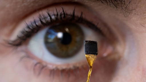
‘Although these mini-hearts are no bigger than a chia seed, they provide us with a powerful platform to discover new treatments more quickly and effectively. Thanks to this technology, we can screen promising compounds at an early stage, which significantly accelerates the development of medicines,’ says Professor Hudson of the Cardiac Bioengineering Lab. In the video below, the scientist talks about his development and the breakthrough.
Realistic representation of disease progression
In the models, the researchers saw that the genetic abnormalities led to fibrosis and reduced pumping function, a realistic representation of disease progression in patients. The team then tested a bromodomain and extra-terminal protein inhibitor, a new class of drugs, which improved heart function in the organoid model.
The Murdoch Children's Research Institute (MCRI) and the Royal Children's Hospital also made important contributions, including through advanced genetic and protein profiling techniques and the use of heart tissue samples from the Melbourne Children's Heart Tissue Bank.
According to Associate Professor Richard Mills (MCRI), the research is an important step towards precision medicine for young heart patients: "This method enables us to model hereditary heart disease in children more accurately and to search specifically for effective therapies. The collaboration with QIMR and the Royal Children's Hospital accelerates this development and opens the door to broader applications in various paediatric heart diseases."
Similar innovations
In 2020, researchers at the LUMC succeeded in developing a 3D mini-heart with adult beating heart cells, made from stem cells in combination with fibroblasts and endothelial cells from the same person. By introducing fibroblasts from patients with arrhythmogenic cardiomyopathy (ACM), the mini-heart exhibited the same disease phenotypes, demonstrating that these connective tissue cells cause the disease. This innovative model makes it possible to identify “perpetrator” and “victim” cells, with the fibroblast as the culprit and the cardiomyocyte as the victim. This model opened doors to regenerative medicine and targeted gene therapy for heart muscle diseases.
In 2022, a researcher developed a so-called fluidic circuit board, a liquid circuit with a standard microfluidic connection, which eliminates the need for flexible tubes in heart-on-a-chip systems. This innovation simplified microfluidic interfacing between components and made the cultivation of heart cells and endothelial cells on chip platforms more reliable, increasing the validity and reproducibility of preclinical pharmaceutical testing with organ-on-a-chip models.


