Researchers at Texas Tech University have developed lifelike 3D-printed tumor models that can serve as an alternative to animal testing when testing surgical imaging technologies. These so-called phantom models are designed to mimic the physical and optical properties of human tumors as accurately as possible. The models are constructed from biologically relevant substances such as lipids, hemoglobin, enzymes, tumor cells, and gelatin, and are printed in a variety of tumor shapes.
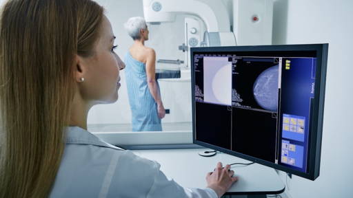
The research is led by Indrajit Srivastava, assistant professor of mechanical engineering, in collaboration with a multidisciplinary team of PhD students and undergraduates. According to Srivastava, these models can be useful in testing afterglow imaging. This is an innovative imaging technique in which light remains present in tumor tissue for longer, up to 10 minutes. Moreover, it also penetrates deeper than traditional fluorescence methods. This gives surgeons more time and precision in locating and removing tumors during surgery.
“Mice are often not a suitable model for this type of research because their skin is too thin to properly mimic human conditions. Our models are designed with layers similar to those of human anatomy, allowing us to test more realistically,” explains Srivastava.
Affordable, scalable, and clinically relevant
An important advantage of the 3D phantom models is their reusability and scalability. Unlike animal testing, researchers can use these models more often, which increases the reliability of research results and reduces costs. Srivastava emphasizes that the models can help bridge the gap between laboratory research and clinical application by providing a more human-centered testing environment.
Data was collected over a period of 18 months within the project. The resulting report was published in the journal ACS Nano. In it, the team describes how the models can be used to test the effectiveness of NIR I/II nanoparticles for use in fluorescence-based surgery. The publication has already led to collaborations with Rice University and the University of Texas at San Antonio, among others.
Optimization of binding agent and models
Srivastava is currently working on optimizing the binding agent in the models so that they are more resistant to temperature fluctuations during transport. In addition, his lab is investigating how tumor cells can survive longer in the phantom model, so that imaging becomes even more realistic and optical signals correspond even better to clinical situations.
These developments are in line with the increasing demand from regulatory bodies such as the FDA and NIH for human-centric research models. “Ultimately, it's about how well a technology translates to the operating room. Our models bring us a big step closer to that goal,” says Srivastava.
3D technology in medicine
The St. Antonius Hospital in Utrecht (Netherlands) has had a 3D lab for some time now, which makes it possible to perform operations better, more accurately, and in less time. The 3D lab creates various types of 3D models that help medical specialists in their work. The models also help patients understand their condition, treatment options, and the possible outcomes of treatment.
A few years ago, researchers at Utrecht University used a 3D printer to create accurate kidney tubules from ‘stable ink’ consisting of gelatin and alginate, which realistically mimic real kidney tissue. These models contain microchannels and form a 3D environment in which kidney cells exhibit naturally diverse behavior, unlike traditional 2D cell cultures. This discovery has contributed to a reduction in the need for animal testing.