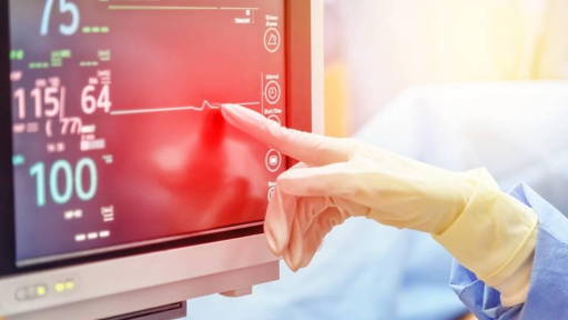
In a groundbreaking advancement in cardiology and medical imaging, Amsterdam UMC has successfully treated a complex cardiac arrhythmia — ventricular arrhythmia — using real-time MRI guidance. This marks a global first, according to the center. The patient underwent a successful ablation procedure while inside an MRI scanner, representing a major leap in precision, safety, and clinical outcomes.
While ablations have traditionally relied on 2D X-ray imaging, MRI now offers a real-time, three-dimensional view of the heart and surrounding tissues. “MRI allows us to track both heart tissue and treatment tools in far greater detail during the procedure. We can instantly see the effect of the ablation on the tissue — a revolution in precision medicine,” explains Dr. Marco Götte, imaging cardiologist and project leader of the MRI intervention program.
Benefits of MRI
MRI-guided ablation shows clear advantages in treating ventricular arrhythmias, where heart chambers contract dangerously fast and irregularly. Standard techniques lack accuracy and tissue visibility — challenges that MRI overcomes.
In recent years, MRI technology has also been used for ablation procedures in other hospitals. For instance, in late 2022, Maastricht UMC+ successfully performed an ablation treatment for cardiac arrhythmias while the patient was inside an MRI scanner — claiming to be the first in the world to do so. This innovation was made possible by advanced software and specialized catheters that enabled physicians to visualize electrical signals directly on 3D MRI images, significantly enhancing the precision of the treatment. In 2023, researcher Ron Holtackers received a Veni grant from the Dutch Research Council (NWO) for his study on ablation therapy within an MRI scanner.
Years of collaboration
This milestone at Amsterdam UMC is the result of years of interdisciplinary collaboration between the departments of Cardiology, Radiology & Nuclear Medicine, Anesthesiology, and Medical Technology. “We have literally brought together the worlds of cardiac MRI and interventional procedures. That requires adjustments to existing workflows, intensive training, and technological innovation,” says Götte.
Prior to the procedure, the team traveled to the United States to practice on pig hearts under controlled conditions. “It’s a strong example of how medical technology, expertise, and thorough preparation come together to shape the future of patient care,” he emphasizes.
MRI intervention suite
The success lays the foundation for a new era of cardiac interventions. A dedicated MRI intervention room with state-of-the-art scanners and MRI-compatible tools will soon open at Amsterdam UMC Heart Center. “Our next milestone is in sight: MRI-guided cardiac tissue biopsies,” says Prof. Dr. Cor Allaart, electrophysiologist.
Since an MRI scanner is a powerful magnet, conventional metal-based tools are unusable. “We had to redesign the entire toolkit in partnership with Siemens, IMRICOR, and MiRTLE Medical — from catheters to ECG systems. It was pioneering work on every level,” adds Götte.
Smarter, safer cardiac care
MRI-guided interventions not only offer greater precision but eliminate radiation exposure altogether. This paves the way for safer, patient-friendly, data-driven cardiac care. The development exemplifies the future of healthcare — smarter, more integrated, and powered by close collaboration between people and technology.
With this world-first, Amsterdam UMC positions itself as a frontrunner in MRI-guided heart interventions — a promising step toward personalized, high-tech care for patients with complex arrhythmias.